DNA and RNA Structure: Key Differences and Worked Examples
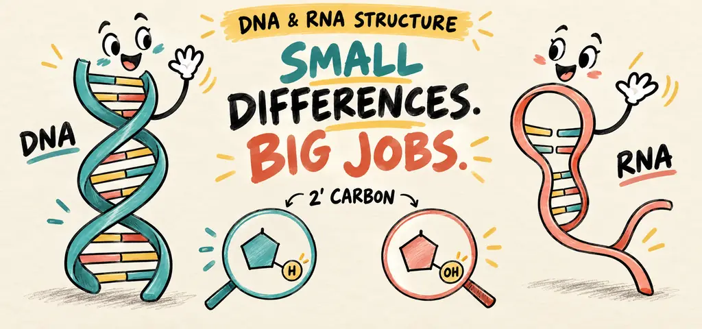
DNA and RNA are chains of nucleotides. Both have a sugar-phosphate backbone and bases. DNA uses deoxyribose and thymine and commonly forms an antiparallel double helix; RNA uses ribose and uracil and is usually single-stranded, with folded regions. Compare the sugar, bases and strand direction before using base percentages to infer structure.
A typical diploid human nucleus is only a few micrometers wide. Before replication, its DNA would stretch roughly two meters. The cell must pack that DNA, expose selected genes for reading, and copy it accurately before division. Start with the nucleotide: its sugar, phosphate, and base explain how a long strand can store information and supply a template for another strand.
Watch the process
DNA and RNA – Part 2
Start With One Nucleotide
A nucleotide is the monomer of a nucleic acid, and it has exactly three parts: one or more phosphate groups, a five-carbon sugar, and a nitrogen-containing base. A nucleotide residue in the usual nucleic-acid backbone contributes one phosphate. The sugar is the hub. Its carbons are numbered \(1’\) through \(5’\), with the prime marks used so that sugar carbons are never confused with the atoms numbered inside the base ring. The base attaches at the \(1’\) carbon. The phosphate attaches at the \(5’\) carbon. The \(3’\) carbon carries a hydroxyl group, and the DNA and RNA polymerases studied here extend a strand by adding a nucleotide to its free \(3’\) hydroxyl.
The sugar is where DNA and RNA differ. Deoxyribose, the sugar in DNA, lacks an oxygen at the \(2’\) carbon; ribose, the sugar in RNA, carries a hydroxyl there. The extra hydroxyl makes RNA more susceptible to backbone hydrolysis, especially in alkaline conditions. Actual RNA lifetime also depends on structure, protective proteins, and degradation machinery; some RNAs are quite stable.
Compare the five standard bases in DNA and RNA. Adenine and guanine are purines, bases built from two fused rings. Cytosine, thymine, and uracil are pyrimidines, bases built from a single ring. DNA uses A, G, C, and T. RNA uses A, G, C, and U, with uracil standing where DNA would carry thymine. Uracil and thymine pair with adenine identically; thymine simply carries an extra methyl group.
Linking Nucleotides Gives the Chain a Direction
Nucleotides join when the phosphate on the \(5’\) carbon of one bonds to the hydroxyl on the \(3’\) carbon of the next. That linkage is a phosphodiester bond, a covalent bond joining two sugars through a phosphate bridge. Repeat it and you get a backbone of alternating sugar and phosphate with the bases hanging off to one side.
Notice what the chemistry has done to the ends. An ordinary linear strand has a \(5’\) end, often bearing phosphate, and a \(3’\) end with a free hydroxyl. End modifications can differ, and circular molecules have no free ends. The two ends are chemically different, so a nucleic acid strand has a direction the way a sentence has a direction. We call the ends the 5’ end and the 3’ end, and by universal convention a sequence written without labels is read left to right from \(5’\) to \(3’\). Label the ends when you work with a sequence so that its direction remains clear.
Two Strands, Paired and Facing Opposite Ways
In a DNA double helix, two strands wind around a common axis with the sugar-phosphate backbones on the outside and the bases stacked in the interior like rungs. The bases pair by hydrogen bonding, and the pairing is specific. Adenine pairs with thymine through two hydrogen bonds. Guanine pairs with cytosine through three. Notice that each pair joins a two-ring purine to a one-ring pyrimidine, so every rung spans the same distance and the helix keeps a constant width. A purine facing a purine would bulge; a pyrimidine facing a pyrimidine would pinch.
The two strands are antiparallel, meaning they run in opposite directions: where one strand runs \(5’\) to \(3’\) left to right, its partner runs \(3’\) to \(5’\) across the same span. Use the labeled ends when predicting replication or writing complementary sequences.
Complementarity has several consequences. First, either strand specifies the other exactly. Given one sequence you can write its partner without any further information, which is what makes accurate copying possible at all and what makes repair possible after damage. Second, G-C-rich duplex DNA often has a higher melting temperature than an A-T-rich duplex of similar length under the same solution conditions. Hydrogen bonding and sequence-dependent base stacking both contribute. Many bacterial origins include readily opened A-T-rich regions, but origin selection and PCR-primer design depend on more than the hydrogen-bond count.
Reading Chargaff’s Rules as Arithmetic
A sample of double-stranded DNA is found to contain 22 percent adenine by base count. Determine the percentages of the other three bases, and decide whether the same reasoning would work on a single-stranded viral genome.
Start from the pairing rule rather than from a memorized formula. Every adenine in a double-stranded molecule is paired with a thymine somewhere on the other strand, so the two totals must be equal. If A is 22 percent, T is also 22 percent.
Now account for the rest. All four bases together must sum to 100 percent, so G and C jointly make up \(100-22-22 = 56\) percent. Guanine pairs only with cytosine, so those two are equal as well, and each accounts for \(56 \div 2 = 28\) percent.
Check the answer against a second relationship. The purines A and G now total \(22 + 28 = 50\) percent, and the pyrimidines T and C total \(22 + 28 = 50\) percent. Purines equal pyrimidines, as they must when every rung of the ladder joins one of each.
A single strand does not impose the same paired-base totals. In a single-stranded genome there is no partner strand forcing A to equal T, so the percentages need not satisfy A=T or G=C, although they still sum to 100 percent. For an accurately measured, pure genome with standard bases, unequal A and T supports a single-stranded model. Equal proportions alone do not prove double-strandedness: a single strand could happen to have matching totals.
Answer
T is 22 percent, G is 28 percent, and C is 28 percent. The A=T and G=C constraints apply to fully complementary double-stranded DNA with standard base pairing; RNA uses U in place of T.
RNA Is the Same Chemistry Used Differently
RNA is a nucleic acid built from ribose nucleotides, usually single-stranded, with lifetimes that depend on RNA type and cellular conditions. Being single-stranded does not mean being shapeless. An RNA strand folds back on itself wherever internal stretches are complementary, producing hairpins, loops, and compact three-dimensional shapes. A folded RNA can therefore have a binding surface and even catalytic activity. The catalytic RNA examples here depend on this folded structure, rather than simply on being single-stranded.
Three RNA classes perform distinct jobs in gene expression.
Messenger RNA (mRNA) is the working copy of a gene’s coding sequence, transcribed from DNA and read by the ribosome. Transfer RNA (tRNA) is the adapter: about seventy to ninety nucleotides folded into a compact shape that carries one specific amino acid at one end and a three-base anticodon at the other. Ribosomal RNA (rRNA) is a structural and catalytic component of the ribosome itself; the bond-forming step of translation is catalyzed by rRNA rather than by protein, which makes the ribosome a ribozyme, an RNA enzyme. Cells also make small regulatory RNAs that bind complementary sequences in messages and block or destabilize them.
What a Base-Composition Table Can and Cannot Establish
A table reports percentages of standard bases in purified genetic material. Assume accurate measurement within rounding and no unusual modified bases. Zero means not detected, not missing data.
| Source | A | T | G | C | U |
|---|---|---|---|---|---|
| Sample 1 | 30.1 | 29.9 | 20.0 | 20.0 | 0 |
| Sample 2 | 24.4 | 24.7 | 25.4 | 25.5 | 0 |
| Sample 3 | 24.7 | 32.8 | 18.5 | 24.0 | 0 |
| Sample 4 | 31.3 | 0 | 24.2 | 17.1 | 27.4 |
Read the structure first. In Samples 1 and 2, A matches T and G matches C to within a fraction of a percent. Those two are consistent with double-stranded DNA, but matching totals do not prove that structure. In Sample 3, A and T differ by eight percentage points and G and C by five and a half, which is inconsistent with fully complementary double-stranded DNA under the stated assumptions. Sample 3 supports a single-stranded DNA model.
Sample 4 contains uracil and no detectable thymine. Under the standard-base assumptions, that supports RNA. Its unequal G and C percentages are inconsistent with a fully complementary double-stranded genome. Merely leaving T blank would not have established RNA; the measured U column supplies the missing evidence.
Now name what the table does not show. It does not identify the organism, because base composition varies widely within every group. It does not report gene content, sequence, or expression. Two samples with identical composition can have completely different sequences, since composition counts bases without regard to order.
Interpreting the result
Samples 1 and 2 are consistent with double-stranded DNA; Sample 3 supports single-stranded DNA, and Sample 4 supports single-stranded RNA under the stated assumptions. Composition constrains structure; it says nothing about sequence or function.
Packing Two Meters Into a Nucleus
A eukaryotic chromosome is not naked DNA. The double helix wraps roughly twice around a core of eight histone proteins, positively charged proteins that bind the negatively charged phosphate backbone by electrostatic attraction. Each DNA-plus-histone unit is a nucleosome, and a string of nucleosomes coils and folds into chromatin, the DNA-protein complex that makes up a chromosome. Loosely packed chromatin is generally more accessible to transcription machinery than tightly packed chromatin. Chromatin packing also regulates access to genes.
Many bacteria store their main genome as a single circular chromosome in a nucleoid region with no surrounding membrane, and many carry small extra circles of DNA called plasmids, which replicate independently and often carry accessory genes, including antibiotic-resistance genes. Some prokaryotes have multiple or linear chromosomes, and not all plasmids are circular. Researchers also use plasmids as vehicles for introducing a gene into a bacterial cell.
Base Structure, Pairing, and Strand Ends
Ring structure classifies bases as purines or pyrimidines; complementary bonding determines which bases pair. A and G are purines because of ring count, and that has nothing to do with which base each pairs with; A pairs with T, and G pairs with C. A student who reasons “A and G are both purines, so they must pair” has merged a structural category with a bonding rule. Say the two rules out loud in sequence and the merge disappears: purines have two rings, pyrimidines have one, and every pair contains one of each.
The labels \(5’\) and \(3’\) identify carbon positions in the sugar. They are not positions in a gene, not distances along a chromosome, and not the order in which bases were discovered. They are carbon numbers on the sugar. A \(5’\) end is the end with no preceding nucleotide joined through its 5′ position; a \(3’\) end ordinarily carries a free 3′ hydroxyl. Once you hold that, “polymerases build only \(5’\) to \(3’\)” stops being a rule to memorize and becomes a description of which chemical group is available.
A nucleic acid is one repeating unit, linked in one direction, with bases whose pairing is fixed by ring size and hydrogen bonding.
Change the sugar and you change stability and lifetime; change the base pairing and you lose the ability of either strand to specify the other.
Label the ends before writing a complementary sequence. A correct base sequence written backward describes a different strand.
Nucleic acid structure
Practice question 1
A sample of double-stranded DNA contains 18 percent cytosine. The percentage of adenine in the sample is
-
18 percent
-
32 percent
-
36 percent
-
64 percent
Practice question 2
Which chemical feature allows ordinary DNA and RNA polymerases to extend a growing strand?
-
the identity of the nitrogenous base, since purines and pyrimidines are added by different enzymes
-
the free 3’ hydroxyl on the sugar, to which these polymerases add the incoming nucleotide
-
the number of hydrogen bonds the base can form with its partner
-
the presence of a methyl group on thymine but not on uracil
Practice question 3
Assume an accurately measured pure genome containing only standard DNA bases. A researcher reports 31 percent adenine, 19 percent thymine, 24 percent guanine, and 26 percent cytosine. The most reasonable conclusion is that the genome is
-
double-stranded DNA, because all four DNA bases are present
-
single-stranded, because A does not equal T and G does not equal C
-
RNA, because thymine is the least abundant base and is being replaced by uracil
-
double-stranded DNA that was measured carelessly, because Chargaff’s rules hold for every nucleic acid
Practice answer key
1. B; 2. B; 3. B.
Practice answer explanations
-
Nucleic acid structure, Question 1. Choice B is correct. In double-stranded DNA every C is paired with a G, so C and G are each 18 percent and together account for 36 percent. The remaining 64 percent is split equally between A and T, giving 32 percent adenine. Choice A applies the pairing rule to the wrong partner by setting A equal to C. Choice C reports the combined G-plus-C total rather than adenine alone. Choice D reports the combined A-plus-T total and forgets to divide it between the two bases.
-
Nucleic acid structure, Question 2. Choice B is correct. The polymerases described here add each incoming nucleotide to the growing strand’s free \(3’\) hydroxyl, so the availability of that group at one end and not the other is what gives a strand its direction of growth. Choice A incorrectly assigns ordinary strand extension to separate purine-adding and pyrimidine-adding polymerases. Choice C confuses the strength of base pairing with the chemistry of the backbone. Choice D names a real difference between thymine and uracil that has nothing to do with the direction of synthesis.
-
Nucleic acid structure, Question 3. Choice B is correct. Chargaff’s equalities hold only when every base has a partner on a complementary strand, so a sample in which A does not equal T and G does not equal C is inconsistent with a fully complementary double-stranded genome under the stated measurement and standard-base assumptions. Choice A treats the presence of all four DNA bases as proof of a double helix, which it is not. Choice C ignores the reported thymine: the reported standard-base genome contains thymine rather than a measured uracil substitution; low thymine alone does not identify RNA. Choice D asserts that the equalities are universal and blames the measurement, which is exactly the assumption the data overturn.
Continue your review at the AP Biology study hub.
Further reading
Review nucleotide chemistry, antiparallel strands and chromosome packaging in OpenStax Biology 2e: DNA Structure and Sequencing.

Related to This Article
More math articles
- West Virginia Algebra 1 Free Worksheets: Printable Algebra 1 Practice with Answer Keys
- Executive agreements
- What Are the Applications of Inverse Trigonometric Functions?
- Information subsidies and venue shopping
- Free Grade 8 English Worksheets for Vermont Students
- Algebra 2 Practice Test
- Touchscreen Monitors for Online Teaching
- Top 10 Free Websites for SHSAT Math Preparation
- Full-Length AFOQT Math Practice Test-Answers and Explanations
- The Best Grade 4 Math Book for New Jersey Students






What people say about "DNA and RNA Structure: Differences and Worked Examples"?
No one replied yet.