Surface Area to Volume Ratio: Cell Size Worked Examples
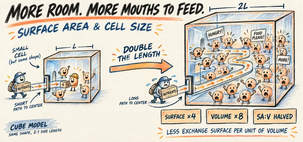
Surface-area-to-volume ratio decreases as a cell grows without changing shape: SA/V = 6/s for a cube and 3/r for a sphere. A larger cell has more total surface, but less surface per unit volume and longer internal paths. Exchange rates also depend on gradients, membrane properties and metabolic demand.
A Calculation Method You Can Check
- Identify the shape and the stated length. A sphere’s radius is half its diameter.
- Calculate surface area and volume using the same length unit.
- Divide area by volume. The quotient has inverse-length units, such as μm⁻¹.
- For similar shapes scaled by factor k, area changes by k², volume by k³ and SA/V by 1/k.
- State the biological assumptions before predicting exchange: compare permeability, concentration gradients and demand per unit volume.
Cut a potato into small pieces and you expose more surface without changing its total volume. Heat has a shorter distance to travel into each piece. Cell size involves a related geometric constraint: materials cross a surface to supply an interior.
Watch the process
Why Are Cells Small?
A cell takes in nutrients and expels waste across its surface, while metabolic demand depends on the material inside its volume. As a cell grows without changing shape, volume increases faster than surface area. Calculate that difference before deciding how size affects exchange.
Check the size units before comparing cells. A typical bacterium is about 1 to 5 micrometers (\(\mu\text{m}\)) across, a typical animal cell 10 to 30, a plant cell often 50 to 100. A micrometer is \(10^{-6}\) meters. A ribosome is measured in nanometers, \(10^{-9}\) meters, and so are membrane thicknesses: a plasma membrane is roughly 7 to 8 nanometers thick. Use these sizes as approximate reference points. When we compare cells of 2 and 6 micrometers on a side, we are talking about an object you would need a good light microscope to see at all.
For a cube of side length \(s\), surface area is \(6s^2\) and volume is \(s^3\). The ratio is therefore \[\frac{\text{SA}}{\text{V}} = \frac{6s^2}{s^3} = \frac{6}{s}.\] Every term but \(s\) cancels, which makes the conclusion unmistakable: the ratio falls as the cell grows, and it falls in inverse proportion to length. One bookkeeping note so the numbers do not confuse you later. Area is measured in square micrometers and volume in cubic micrometers, so the ratio carries units of \(1/\mu\text{m}\) rather than being a pure number. AP items report it as a ratio such as \(3:1\) and expect you to compare two cells measured the same way, which is all the comparison needs. For a sphere the algebra gives \(3/r\), a different constant but the same behavior. What matters is not the constant. It is that the exponent on volume is one larger than the exponent on area.
A Worked SA:V Calculation
A cube-shaped cell has a side length of 2 micrometers. A second cube-shaped cell of the same material has a side length of 6 micrometers. Compare their surface areas, volumes, and surface-area-to-volume ratios, and state what the comparison predicts about exchange.
Take the small cell first. Surface area is \(6s^2 = 6(2)^2 = 24\ \mu\text{m}^2\). Volume is \(s^3 = (2)^3 = 8\ \mu\text{m}^3\). The ratio is \(24/8 = 3\), or \(3:1\).
Now the large cell. Surface area is \(6(6)^2 = 216\ \mu\text{m}^2\). Volume is \((6)^3 = 216\ \mu\text{m}^3\). The ratio is \(216/216 = 1\), or \(1:1\).
Read the two results against each other. Tripling the side length multiplied surface area by 9 and volume by 27, so the ratio fell by a factor of 3, exactly as \(6/s\) predicts. If both cells have the same demand per unit volume and exchange capacity per unit area, the larger cell has 27 times the demand and only 9 times the delivery surface, so each unit of its cytoplasm is served by one third as much membrane. Diffusion distance to the center has also tripled, which compounds the problem.
Answer
Small cell: \(24\ \mu\text{m}^2\), \(8\ \mu\text{m}^3\), ratio \(3:1\). Large cell: \(216\ \mu\text{m}^2\), \(216\ \mu\text{m}^3\), ratio \(1:1\). The larger cell has one third the exchange surface per unit of cytoplasm. Under matched exchange and metabolic conditions, that creates a greater supply constraint; geometry alone does not measure actual uptake or prove cell failure.
The Same Calculation for a Sphere
A sphere provides another useful idealized cell shape. Its formulas are \(\text{SA} = 4\pi r^2\) and \(V = \tfrac{4}{3}\pi r^3\). Compute the surface-area-to-volume ratio for a spherical cell of radius \(3\ \mu\text{m}\) and for one of radius \(6\ \mu\text{m}\), and state the general rule the two results demonstrate.
Take the small sphere. Surface area is \(4\pi(3)^2 = 4\pi(9) = 36\pi \approx 113.1\ \mu\text{m}^2\). Volume is \(\tfrac{4}{3}\pi(3)^3 = \tfrac{4}{3}\pi(27) = 36\pi \approx 113.1\ \mu\text{m}^3\). The ratio is \(113.1 / 113.1 = 1.0\), or \(1:1\).
Now the large sphere. Surface area is \(4\pi(6)^2 = 4\pi(36) = 144\pi \approx 452.4\ \mu\text{m}^2\). Volume is \(\tfrac{4}{3}\pi(6)^3 = \tfrac{4}{3}\pi(216) = 288\pi \approx 904.8\ \mu\text{m}^3\). The ratio is \(452.4 / 904.8 = 0.5\), or \(1:2\).
Now derive the shortcut so you never have to do that arithmetic again under time pressure. Divide the two formulas symbolically: \[\frac{\text{SA}}{V} = \frac{4\pi r^2}{\tfrac{4}{3}\pi r^3} = 4\pi r^2 \cdot \frac{3}{4\pi r^3} = \frac{3}{r}.\] The \(4\), the \(\pi\), and two powers of \(r\) all cancel, leaving \(3/r\). Check it against the numbers: \(3/3 = 1.0\) and \(3/6 = 0.5\). They match.
Read the two shortcuts side by side. A cube gives \(6/s\) and a sphere gives \(3/r\). The constants differ because the shapes differ, but the form is identical: the ratio is a constant divided by one length. Scale every length by the same factor while keeping shape unchanged, and the ratio decreases by that factor.
Answer
Small sphere: \(36\pi \approx 113.1\ \mu\text{m}^2\), \(36\pi \approx 113.1\ \mu\text{m}^3\), ratio \(1:1\). Large sphere: \(144\pi \approx 452.4\ \mu\text{m}^2\), \(288\pi \approx 904.8\ \mu\text{m}^3\), ratio \(1:2\). In general \(\text{SA}/V = 3/r\), so doubling the radius halves the ratio.
Cells answer this constraint in four ways, and each one is a familiar structure once you see the geometry. They stay small and divide, which is why bacteria and most eukaryotic cells are microscopic. They flatten or elongate, because a shape far from spherical has more surface for the same volume, which is what a red blood cell’s biconcave disc and a neuron’s axon accomplish. They fold the exchange surface itself: microvilli on an intestinal cell, root hairs on a plant root, alveoli in a lung, cristae inside a mitochondrion. And they move material by bulk flow rather than waiting for diffusion, using cytoplasmic streaming or a circulatory system.
Why the Ratio Predicts a Rate, Not Just a Shape
A biology class models cell size using cubes of agar containing an indicator dye that changes color when acid soaks in. They cut cubes of side 1 cm, 2 cm, and 3 cm, drop all three into acid for the same ten minutes, then slice each cube open and measure how deep the color change went. The depth of penetration is about the same in all three cubes: roughly 4 mm from every face. What should they conclude, and what does the ratio add that the penetration depth alone does not say?
Start with the equal penetration depths. The observed fronts penetrated the same distance in the same time under matched conditions. That supports similar local penetration behavior, but it does not measure the total number of molecules entering each differently sized cube.
What differs is the fraction of each cube that got served. The 1 cm cube has volume \(1\ \text{cm}^3\); a 4 mm rind leaves an untreated core with side \(1.0-0.8=0.2\) cm and volume \(0.2^3=0.008\) cubic centimeters, so 99.2 percent is treated. The 3 cm cube has volume \(27\ \text{cm}^3\), and an untreated core of side \(3-0.8 = 2.2\) cm has volume \(2.2^3 \approx 10.6\ \text{cm}^3\), so about 39 percent of the block remains unchanged at the ten-minute observation. This does not mean that region can never be reached. The ratio makes the same point in one number: \(6/1 = 6\) for the small cube and \(6/3 = 2\) for the large one.
The ratio identifies less exchange surface per unit volume in a larger, similarly shaped object. With the same penetration depth, a smaller fraction changes color in the larger cube. Actual uptake also depends on permeability, gradients and time; the ratio alone does not establish a rate or a biological failure threshold.
Answer
The penetration distance after ten minutes is similar in all three cubes. At that measured depth, the changed fraction falls as cube size rises. SA/V describes the geometry; the changed fraction requires the depth measurement too.
Check All Three Agar Cubes
For the stated ten-minute example, 4 mm equals 0.4 cm. The unchanged core has side max(s − 2d, 0), because penetration occurs from both opposing faces. Changed fraction = 1 − [max(s − 2d, 0)/s]³. This idealized calculation assumes a uniform front from every face.
| Cube side (cm) | SA/V (cm⁻¹) | Core side (cm) | Changed volume (%) |
|---|---|---|---|
| 1 | 6 | 0.2 | 99.2 |
| 2 | 3 | 1.2 | 78.4 |
| 3 | 2 | 2.2 | 60.6 |
The larger cube can have more changed volume in absolute terms while a smaller percentage has changed. An indicator front is a model observation, not a direct measurement of membrane permeability or total molecular uptake by a living cell.
The Confusion to Clear Up Here
Surface area and surface-area-to-volume ratio can change in different directions. A growing cell gains surface area. It gains volume faster. So the absolute amount of membrane goes up while the amount of membrane serving each unit of cytoplasm goes down. If a stem asks what happens to surface area as a cell grows, the answer is that it increases. If it asks what happens to the ratio, the answer is that it decreases. Read which quantity the question named.
Increasing cell size creates both an exchange constraint and a diffusion-distance constraint. One is supply: not enough membrane per unit of volume. The other is distance: the center of a large cell is farther from the surface, and in a simple diffusion model with the same diffusion coefficient, characteristic time scales with distance squared. Doubling a comparable distance therefore gives a roughly fourfold characteristic time, not a fixed arrival time for every molecule. Microvilli increase exchange area; they do not by themselves shorten the path from the plasma membrane to the center of the cell. Keep the absolute area and the ratio separate.
Review: Surface Area, Volume, and the Limits on Cell Size
Surface grows with the square of a length while volume grows with the cube, so the ratio falls when a cell grows without changing its overall shape.
Change the cell’s size or shape and you change how much exchange surface serves each unit of cytoplasm. That exchange capacity places an important constraint on sustainable metabolic demand; transporters, internal transport, and organelle activity also matter.
Do not memorize \(6/s\) and \(3/r\) as separate facts. The same scaling relationship applies when a shape is unfamiliar.
Apply the ratio to the metabolically active volume. The ratio does not set a single universal maximum size. Large eggs can contain substantial stored yolk, and some large plant or algal cells contain a large vacuole with a comparatively thin layer of cytoplasm. Such organization, internal transport and specialized exchange surfaces help explain why total size alone is not enough. Metabolically active volume and local transport paths matter; no single adjusted ratio predicts every cell’s limit.
Surface area, volume, and exchange
Practice question 1
A spherical cell doubles its radius. Its surface-area-to-volume ratio
-
doubles
-
is unchanged
-
quadruples
-
is halved
Practice question 2
A cube-shaped cell with a side length of 3 units has a surface-area-to-volume ratio of
-
\(1:1\)
-
\(2:1\)
-
\(3:1\)
-
\(6:1\)
Practice question 3
An intestinal cell is covered in microvilli. The best explanation for this structure is that microvilli
-
reduce the cell’s volume so less nutrient is required
-
allow the cell to exceed the limits imposed by diffusion inside the cytoplasm
-
increase absorptive surface area without a proportional increase in cell volume
-
convert active transport into simple diffusion
Practice answer key
1. D; 2. B; 3. C.
Practice answer explanations
-
Surface area, volume, and exchange, Question 1. Choice D is correct. For a sphere the ratio is \(3/r\), so doubling the radius halves the ratio. Choice A inverts the relationship. Choice B claims the ratio is scale-independent, which contradicts the unequal exponents on area and volume. Choice C multiplies where the relationship divides.
-
Surface area, volume, and exchange, Question 2. Choice B is correct. Surface area is \(6(3)^2 = 54\) and volume is \(3^3 = 27\), giving \(54/27 = 2\), or \(2:1\); the shortcut \(6/s\) gives the same result. Choice A corresponds to a side length of 6. Choice C corresponds to a side length of 2. Choice D corresponds to a side length of 1, the unit cube.
-
Surface area, volume, and exchange, Question 3. Choice C is correct. Microvilli are folds that multiply absorptive surface while adding little volume, which raises the ratio. Choice A misreads folding as volume reduction. Choice B overclaims, since folding the outer surface does nothing about diffusion distance within the cytoplasm. Choice D confuses the amount of surface with the mechanism of transport across it.
Continue your review at the AP Biology study hub.
Connect Geometry to Membrane Function
Review membrane structure and fluidity to separate movement within a membrane from passage across it. Then use channels, carriers and saturation to see why exchange capacity is more than surface area alone.
Sources for Further Study
OpenStax explains cell size and surface-area-to-volume scaling. Practical Biology describes an agar-cube diffusion investigation; the numbers above belong to this lesson’s stated example.

Related to This Article
More math articles
- Wisconsin FORWARD Grade 4 Math Free Worksheets: Free Printable PDFs Covering Every Grade 4 Skill
- Algebra Puzzle – Challenge 56
- Advanced Vocabulary Words: Gauche to Illustrious
- Time Travel Adventure: How to Perform Indirect Measurement in Similar Figures
- Graphs and Data Analysis for 5th Grade: Bar, Line, and Circle Graphs
- Advanced Vocabulary Words: Fantastic to Garrison
- Baccarat Scoreboards: What the Big Eye Road Actually Tracks
- The Best Algebra 1 Book for New Jersey Students
- Horse Racing Odds Explained: Probability & Expected Value
- Customary Unit Conversions Involving Mixed Numbers and Fractions









What people say about "Surface Area to Volume Ratio: Cell Size Worked Examples"?
No one replied yet.