Endosymbiotic Theory: Organelles, Evidence and Gene Transfer
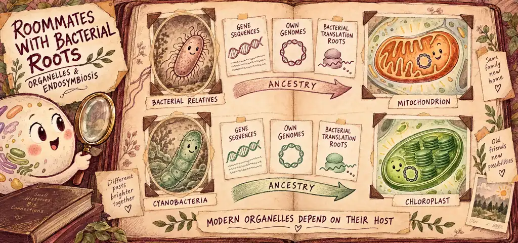
Endosymbiotic theory explains the bacterial ancestry of mitochondria and chloroplasts. Separate genomes, sequence relationships, bacterial-derived translation machinery and division of existing organelles support that history. A double membrane is supporting evidence; it does not establish ancestry by itself. Compartments also serve present-day functions by separating reactions and maintaining local conditions.
Separate Function from Ancestry
- For function, ask what a membrane separates: pH, substrates, enzymes or an electrochemical gradient.
- For protein orientation, follow the cytosolic and lumen-facing surfaces through each budding and fusion event.
- For ancestry, compare genetic sequences and translation machinery; count membranes as one supporting observation.
- For gene transfer, distinguish nuclear location from evolutionary origin. A host-encoded protein need not have come from the ancestral bacterium.
A lysosome maintains an acidic interior while the surrounding cytosol remains near neutral. Its membrane helps keep those conditions separate. Internal membranes also let a cell concentrate enzymes and substrates in one place or contain potentially harmful intermediates.
Watch the process
Endosymbiotic Theory
A membrane-bounded compartment can maintain a local environment suited to its reactions. Lysosomes contain digestive enzymes; peroxisomes compartmentalize reactions involving hydrogen peroxide. The inner mitochondrial membrane has another role: it limits proton movement, allowing an electrochemical gradient to drive ATP synthesis.
Compartmentalization also increases the membrane surface available inside a cell. A folded internal membrane adds enormous surface without adding much volume. The cristae of a mitochondrion, the thylakoid stacks of a chloroplast, and the sheets of rough ER are all the same trick: pack more working surface into a fixed space.
Compartments can also change how often reactants encounter one another. Reaction rate depends on how often the reactants meet. In an idealized calculation, moving the same numbers of enzyme and substrate molecules from a cell-sized volume into a compartment one thousandth as large would increase each concentration a thousandfold. Real compartments do not automatically achieve that enrichment; molecule numbers, transport and reactions must be considered. Compartmentalization is therefore a way of increasing encounter frequency without making additional reactants. The actual rate also depends on enzyme saturation and transport across the boundary, and maintaining a compartment can cost energy.
The Endomembrane System as One Connected Whole
Do not read the organelles of the endomembrane system as separate objects. The nuclear envelope, the endoplasmic reticulum, the Golgi apparatus, vesicles, lysosomes, vacuoles, and the plasma membrane form a single trafficking network whose parts exchange membrane with one another. The outer nuclear membrane is physically continuous with the ER. Much of the traffic between other parts of this network uses transport vesicles, small membrane sacs that bud off one compartment and fuse with another, so membrane and cargo travel together.
Vesicle traffic changes membrane area while preserving membrane orientation. Whenever a vesicle buds, the compartment it leaves loses a patch of membrane; whenever a vesicle fuses, the compartment it joins gains one. The cell is therefore constantly moving membrane area between compartments, and the orientation is preserved: the face of the membrane that pointed into the ER lumen ends up pointing out of the cell after exocytosis. This is why a protein that is glycosylated in the ER lumen ends up with its sugar chain on the outside of the cell surface, and it is why the two faces of every membrane in the system stay chemically different.
Tracing a Membrane Protein to Its Final Orientation
A membrane protein is synthesized on the rough ER. One region of the finished protein carries a carbohydrate chain, and that chain is added while the region sits in the ER lumen. After the protein reaches the plasma membrane, will the carbohydrate face the cytosol or the outside of the cell? Explain the reasoning rather than recalling the answer.
Work the route one fusion event at a time and keep track of which side is which. The protein is inserted into the ER membrane, and the carbohydrate-bearing region is in the ER lumen. A vesicle buds from the ER; the old lumen becomes the vesicle interior, so the sugar now faces the inside of the vesicle. The vesicle fuses with the Golgi, and the vesicle interior becomes the Golgi lumen, so the sugar still faces a lumen. Another vesicle buds from the Golgi and carries the protein to the plasma membrane.
Now the last step, which is the one that flips your intuition without flipping the molecule. When the vesicle fuses with the plasma membrane, the vesicle interior opens to the outside of the cell. Nothing about the protein rotated. The compartment it was facing simply became the extracellular space.
Answer
The carbohydrate faces outward, into the extracellular space, because the lumen-facing surface keeps its orientation through budding and fusion. Exocytosis exposes it to the extracellular space. Topological equivalence does not mean every compartment is continuously physically connected.
Where Two Organelles Came From
Endosymbiotic theory holds that mitochondria and chloroplasts descend from free-living prokaryotes that were engulfed by an ancestral host cell and retained rather than digested. Evaluate the endosymbiotic explanation against structural and genetic evidence. Four lines of evidence support it. Consider what each observation supports and what it cannot establish alone.
First, mitochondria and primary chloroplasts have a double membrane, a feature consistent with an endosymbiotic history. The familiar engulfment drawing is a simplified model, not proof of the exact ancestry of each modern membrane. Membrane number alone cannot establish origin: the nuclear envelope also has two membranes.
Second, both retain their own genomes, separate from the nuclear genome. These are often represented as circular DNA, a common bacterial feature, although organelle genome arrangements vary. More decisive evidence comes from sequence comparisons linking mitochondrial genes to bacterial relatives and chloroplast genes to cyanobacteria.
Third, both have ribosomes descended from bacterial ribosomes and distinct from the host’s cytosolic 80S ribosomes. Chloroplast ribosomes are typically 70S; mitochondrial ribosomes vary, and mammalian mitochondrial ribosomes are 55S. The S value describes sedimentation in a centrifuge and is not a mass to add arithmetically. Certain antibiotics inhibit bacterial and organellar translation while sparing cytosolic translation, but sensitivity depends on the drug and organelle. The important evidence is shared molecular ancestry, not a universal 70S label.
Fourth, both multiply from preexisting organelles by division that resembles bacterial binary fission, although host proteins regulate the process. They do not arise through mitosis of the host nucleus. In the usual cellular cycle, mitochondria grow and divide from preexisting mitochondria, using imported host-encoded components as well as products of their own genome. Growth and division support continuity; they do not make the organelle genetically self-sufficient.
The organelles are not independent organisms now. Many genes of endosymbiont origin were transferred to the nuclear genome or lost, and modern organelles depend on numerous nuclear-encoded proteins translated in the cytosol and imported. Not every nuclear-encoded mitochondrial protein came from the endosymbiont; host pathways also contributed proteins. Sequence evidence can distinguish these histories.
Building the Argument (Science Practice 6)
A student is given three observations about mitochondria: they have two membranes, they contain circular DNA, and an antibiotic known to bind bacterial and mitochondrial ribosomes reduces the synthesis of certain mitochondrial proteins without affecting cytosolic protein synthesis. Construct a claim with evidence and reasoning.
Claim. The mitochondrion descends from a free-living bacterium that was taken up by an ancestral host cell.
Evidence. Two membranes, a circular genome, and organellar translation sensitive to a bacterial-ribosome antibiotic while the cytosol’s ribosomes are not.
Reasoning. The observations together support bacterial ancestry. Separate genomes and bacterial-like translation machinery are especially informative. Double membranes are consistent with endosymbiosis but are not unique to it, and their exact historical origin cannot be reconstructed from membrane count alone. Gene-sequence comparisons provide a further test. None of these observations makes a modern mitochondrion an independent organism: it relies on many host-encoded proteins.
Conclusion
Together, the separate genome and bacterial-like translation machinery support bacterial ancestry. The double membrane is consistent with that history.
The Confusion to Clear Up Here
Endosymbiosis does not explain the origin of every organelle. For AP Biology, the central cases are mitochondria and chloroplasts. The nucleus and endomembrane system have a different evolutionary history, often illustrated with membrane-remodeling models. The nuclear envelope has two membranes and encloses the main cellular genome; the ER, Golgi, and lysosomes do not contain separate genomes. Do not infer that every double-membrane compartment came from an endosymbiont.
Having its own DNA does not make an organelle an independent organism. A mitochondrion has a genome, but that genome encodes only a small fraction of the proteins the organelle needs. The rest are transcribed from nuclear genes, translated on cytosolic ribosomes, and imported. Modern mitochondria normally depend on the host for sustained growth and reproduction. Their own genome and imported nuclear-encoded proteins work together; neither observation alone makes them independent bacteria or disproves bacterial ancestry.
Review: Compartmentalization and the Endosymbiotic Origin of Organelles
A membrane makes an inside, and an inside can hold conditions and gradients the rest of the cell cannot.
Add or remove a membrane boundary and you change what chemistry the cell can run in parallel.
Do not treat “double membrane” as a label to memorize. A double membrane is consistent with endosymbiosis, but sequence relationships and translation machinery provide stronger ancestry evidence than membrane count alone.
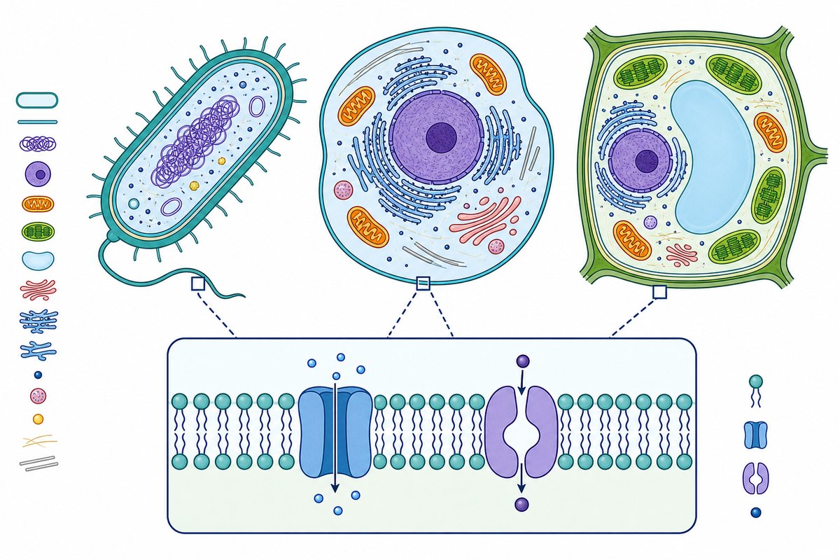
Compartments and endosymbiosis
Practice question 1
Which observation provides the strongest single line of evidence that mitochondria descend from free-living prokaryotes?
-
mitochondria are found in nearly all eukaryotic cells, from single-celled yeasts to mammals
-
a mitochondrion’s inner membrane is deeply folded, which increases its working surface area
-
mitochondria carry out the oxygen-consuming reactions that supply most of the cell’s ATP
-
mitochondrial ribosomal components have bacterial sequence similarities and can be inhibited by certain antibiotics that spare cytosolic translation
Practice question 2
A lysosome has one membrane and a mitochondrion has two. The difference is best explained by the fact that
-
lysosomes are older than mitochondria, so they were built with fewer layers
-
lysosomes are part of endomembrane trafficking, while mitochondria have an endosymbiotic history consistent with two surrounding membranes
-
a mitochondrion is the larger organelle and needs a second membrane for structural support
-
a lysosome lost its outer membrane during evolution
Practice question 3
Most mitochondrial proteins are encoded by nuclear genes. This observation
-
argues against endosymbiosis, because an engulfed cell would have kept its own genes
-
shows that mitochondria are not organelles, since an organelle must be genetically self-sufficient
-
is consistent with transfer of endosymbiont genes to the nuclear genome over evolutionary time
-
indicates that the host cell assembled the mitochondrion from nuclear-encoded parts
Practice answer key
1. D; 2. B; 3. C.
Practice answer explanations
-
Compartments and endosymbiosis, Question 1. Choice D is correct. Bacterial sequence similarities in mitochondrial ribosomal components, together with selective antibiotic sensitivity, support bacterial ancestry. Mitochondrial ribosome sizes vary; the evidence does not require all of them to be 70S. Choice A states a distribution fact that is equally consistent with the host having built the organelle in a common ancestor. Choice B names a folding adaptation that raises surface area, which is a functional advantage the host could equally well have built and therefore says nothing about origin. Choice C names a metabolic function that does not by itself indicate ancestry.
-
Compartments and endosymbiosis, Question 2. Choice B is correct. Lysosomes form and mature through endomembrane trafficking and have one surrounding membrane, whereas mitochondrial double membranes are consistent with an endosymbiotic history. This simplified comparison does not establish the exact ancestry of each membrane; sequence evidence is more decisive. Choice A substitutes relative age for mechanism. Choice C explains membrane number by organelle size, but membrane count records how the compartment arose, not how big it is, and many large compartments such as the central vacuole have one membrane. Choice D asserts a loss for which there is no evidence and which reverses the actual origin.
-
Compartments and endosymbiosis, Question 3. Choice C is correct. Transfer of some endosymbiont genes to the nuclear genome is consistent with endosymbiosis, so nuclear encoding does not refute the theory. Other mitochondrial proteins were recruited from host pathways; the location of a gene alone does not establish its ancestry. Choice A treats the observation as fatal when the theory predicts it, because transfer and loss of redundant genes can occur while the host supplies the necessary products. Choice B redefines organelle around genetic self-sufficiency, a requirement no organelle meets. Choice D confuses where the genes ended up with who built the compartment; nuclear encoding says nothing about the origin of the membranes.
Continue your review at the AP Biology study hub.
Connect Compartments to Membrane Function
Use the fluid mosaic membrane model to review membrane structure and asymmetry. Then compare surface area and cell size to see how working membrane area and internal transport paths constrain a cell.
Sources for Further Study
OpenStax reviews endosymbiotic evidence and eukaryotic origins. The Cell explains the origin and evolution of cells, including organelle genomes and dependence on imported proteins.

Related to This Article
More math articles
- Bail
- Overview of ATI TEAS 7 Mathematics Test
- The Best Grade 6 ELA Practice Tests for Arkansas Students
- GED Testing Accommodations and Support for Students with Disabilities
- How to Use Area Models to Factor Variable Expressions?
- How to Find Midpoint? (+FREE Worksheet!)
- PERT Math Formulas
- New York Grade 6 Math Worksheets: 72 PDF Links
- 3rd Grade WVGSA Math Worksheets: FREE & Printable
- ASTB Math FREE Sample Practice Questions







What people say about "Endosymbiotic Theory: Organelles, Evidence and Gene Transfer"?
No one replied yet.