Cell Cycle Checkpoints: G1, G2 and Spindle Control
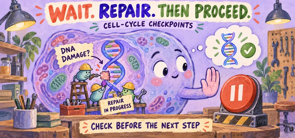
The cell cycle has three major checkpoint controls: G1 control helps decide whether a cell enters DNA synthesis; G2 control delays mitosis when replication or DNA integrity is compromised; the spindle assembly checkpoint delays chromosome separation when kinetochores remain unattached. A checkpoint is a signaling mechanism, not a physical gate.
Skin can replace many damaged cells through division, whereas most mature neurons remain outside the division cycle. These cell types share essentially the same inherited genome but express different proteins and respond differently to growth signals. Cell specialization includes regulation of whether and when division occurs.
Watch the process
Cell cycle control | Cells | MCAT | Khan Academy
A cell prepares for division through growth, DNA replication, and checks on its condition. Mitosis and cytokinesis complete the process. The controls that govern entry into each stage help explain why different cell types divide at different times.
Interphase covers three phases. In G\(_1\) the cell grows, builds organelles, and carries out its ordinary work. In S phase the cell replicates its DNA, so that each chromosome comes to consist of two sister chromatids, two normally matching DNA copies held together by cohesion proteins, especially near their centromeres. A centromere is a chromosome region where a kinetochore forms for spindle attachment; each replicated chromatid has its own centromeric DNA. In G\(_2\) the cell continues to grow, prepares its duplicated centrosomes in animal cells, and synthesizes the proteins that mitosis will need. Only then does M phase, mitosis plus cytokinesis, divide the nucleus and the cytoplasm.
Cells that stop dividing exit from G\(_1\) into G\(_0\), a nondividing state. Most mature neurons stay there permanently, while liver cells can be recruited back into G\(_1\) by growth factors after injury. A cell in G\(_0\) is metabolically busy; it is simply not preparing to divide.
The proportions matter. An example mammalian cell in culture may complete a cycle in roughly 24 hours and spends under an hour in M phase, which is why a random microscope field is mostly interphase nuclei.
Cell-cycle lengths vary widely. For a simplified 24-hour example, G\(_1\) runs about 11 hours, S about 8, G\(_2\) about 4, and M under 1. The consequence for a microscope slide is direct: if roughly 4 percent of the cycle is M phase, then a classroom model that equates phase fractions with time fractions predicts about 4 percent mitotic cells. This approximation assumes representative sampling of cycling cells and ignores unequal age frequencies created by population growth. Most cells in such a sample would therefore appear to be in interphase.
One warning about interphase. It is easy to read “the cell is not dividing” as “the cell is idle,” and the older textbook name for interphase, resting phase, made that worse. An interphase cell is transcribing, translating, respiring, transporting, and in S phase copying its nuclear DNA with proofreading (about six billion base pairs in a typical diploid human cell before replication). Interphase therefore includes substantial cellular activity; mitosis is a distinct redistribution of the replicated chromosomes.
Three Checkpoints
A checkpoint is a control point where the cycle stops unless specific internal and external conditions are met. Follow three major checkpoint controls.
G\(_1\) control includes a growth-dependent restriction point and DNA-damage checkpoint mechanisms. These are related controls rather than identical names for one molecular switch. It asks whether the cell is large enough, whether nutrients are adequate, whether growth-factor signals are present, and whether the DNA is undamaged. A cell that passes it is generally committed to complete the cycle. A cell that does not meet the requirements may arrest or enter G\(_0\).
The G\(_2\) checkpoint helps prevent mitotic entry when replication is incomplete or DNA damage is detected; it does not guarantee that every base is error free. Entering mitosis with half-replicated chromosomes would tear them apart, so this checkpoint holds the cell until replication is verified.
The spindle assembly checkpoint operates at the metaphase-to-anaphase transition. It holds anaphase until every kinetochore, the protein platform built on each chromatid at the centromere where spindle microtubules grip, is attached. Proper alignment requires the two sister kinetochores to connect to opposite poles, not one kinetochore to both poles. An unattached kinetochore emits a signal that blocks the transition, and one unattached chromosome is enough to hold the whole cell. This is the safeguard against distributing chromosomes unevenly.
Cyclins and Cdks: What Operates the Checkpoints
Cyclins and CDKs promote cell-cycle transitions. Checkpoint signaling can restrain these proteins or other regulators when a problem is detected.
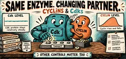
A cyclin-dependent kinase, or Cdk, is a protein kinase. In the simplified cell-cycle model used here, its abundance is relatively stable while its activity changes. A cyclin is a regulatory protein whose concentration rises and falls in a repeating pattern across the cycle, which is where the name comes from. Cyclin binding is required for activation of the cell-cycle Cdks discussed here. Activating or inhibitory phosphorylation and inhibitor proteins also regulate activity, so binding alone need not be sufficient.
Consider mitotic entry at the G\(_2\) checkpoint. Cyclin accumulates gradually through S and G\(_2\). As it accumulates, more of it binds Cdk, forming the complex historically called maturation-promoting factor, or MPF. In this simplified model, sufficient mitotic Cdk activity phosphorylates a set of target proteins that trigger nuclear-envelope breakdown, chromosome condensation, and spindle assembly. The cell enters mitosis. Mitotic cyclin is later targeted for degradation through the anaphase-promoting complex/cyclosome (APC/C), with other regulatory signals controlling the timing. Cyclin degradation lowers mitotic Cdk activity and helps permit mitotic exit. Rising cyclin drives entry and cyclin destruction permits exit. Cyclin accumulation and destruction help produce repeated changes in Cdk activity.
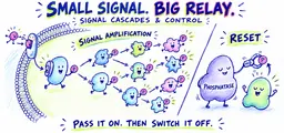
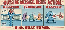
A different cyclin-Cdk pair operates the G\(_1\) checkpoint, and it is the one growth factors talk to. In a common growth-factor signaling model, a receptor tyrosine kinase activates a pathway that changes gene expression, G\(_1\) cyclin is made, G\(_1\) cyclin-Cdk becomes active, and the cell passes the restriction point. This connects growth signaling to cell-cycle control: the decision to divide is the response stage of a signal transduction pathway.
Internal brakes act on the same complexes. When DNA damage is detected, the p53 protein accumulates and activates transcription of a Cdk inhibitor protein, which binds cyclin-Cdk complexes and shuts them off. This response can delay the cycle to allow repair. Persistent damage can instead lead to senescence or apoptosis, depending on the cell and its signaling pathways; these outcomes are not an automatic sequence.
What a Checkpoint Actually Is
A checkpoint is not a physical gate and not a place. It is a signaling system that can delay a transition when a monitored condition is not met. Different checkpoints act on different regulatory targets. Three functional roles help describe a checkpoint, and naming them turns vague checkpoint questions into answerable ones.
A sensor detects the condition, such as a protein complex that binds single-stranded DNA at a break, or a kinetochore that has no microtubule attached. A transducer carries that information to the machinery, usually by phosphorylating something or by changing the concentration of a regulatory protein. An effector produces the arrest. DNA-damage checkpoints can inhibit cyclin-Cdk activity; the spindle checkpoint instead restrains APC activity to delay anaphase.
Read a checkpoint item by asking which of the three the stem broke. If a required sensor is lost, the checkpoint may fail to detect the problem and allow progression. If a required effector is lost, the cell may detect damage yet fail to produce the expected arrest. Both produce the same headline outcome, “the cell divides with damaged DNA,” but they are different lesions, which can be distinguished using measurements such as accumulation of a damage-response protein.
Which Checkpoint Would Catch It?
Four defects are described. For each, name the checkpoint that should detect the problem, and state what would happen if that checkpoint were also disabled.
(a) A cell is starved of growth factor. The G\(_1\) checkpoint is the one that assesses external signals, cell size, and nutrient supply. It holds the cell before S phase, and a cell held long enough exits to G\(_0\). If the G\(_1\) checkpoint were disabled, the cell would enter S phase despite inadequate growth signaling, if other requirements for progression are met. This is one way growth control can be bypassed.
(b) Ultraviolet light produces DNA lesions during G\(_1\). The G\(_1\) checkpoint again, through the p53 pathway. If this arrest mechanism fails, replication may encounter the lesion, stall, or introduce a mutation during bypass. Damage is not automatically converted into a mutation in both daughter molecules, and DNA repair can also occur after replication.
(c) A replication fork stalls and one region of a chromosome is left uncopied. Replication-stress signaling during S phase and checkpoint controls before mitosis help prevent progression with incomplete DNA replication. If the relevant controls fail, mitosis may begin with incompletely replicated DNA, increasing the risk of chromosome bridges, breakage, or unequal inheritance.
(d) One chromosome’s kinetochore fails to capture microtubules. The spindle assembly checkpoint holds the metaphase-to-anaphase transition until every kinetochore is attached. Disable it and anaphase begins before correct attachment, increasing the risk of a lagging chromosome and daughters with extra or missing chromosomes.
Read the pattern across the four. Each checkpoint precedes the irreversible step it protects, and the predicted defect follows from allowing an unprepared cell to attempt the next step.
Answer
(a) and (b) G\(_1\); (c) G\(_2\); (d) spindle assembly. Losing a checkpoint can allow progression despite a problem, increasing the risk of damage or unequal chromosome inheritance.
Review: The Cell Cycle and Its Checkpoints
Cell-cycle controls help coordinate progression with growth, DNA replication, and chromosome attachment; they reduce errors without guaranteeing an error-free division.
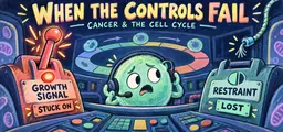
Loss of a checkpoint can permit inappropriate progression. It does not guarantee a faster cycle; other controls, severe damage, or cell death may still stop the cell.
Distinguish loss of a checkpoint brake from activation of an intact checkpoint. Losing the brake can permit inappropriate progression, although other defects or controls may still stop division.
Distinctions to keep clear
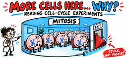
Arrest is not the same as checkpoint failure. A cell that accumulates in G\(_2\) may have a working checkpoint responding to damage, or a separate defect in the machinery required to enter mitosis. A cell with a broken G\(_2\) checkpoint does the opposite: it enters mitosis when it should have stopped. Accumulation identifies where cells are delayed; it does not, by itself, establish why. Inspect measurements of DNA damage, checkpoint signaling, or mitotic Cdk activity.
Distinguish G\(_0\) from a block inside the cycle. A cell in G\(_0\) has exited the cycle, not paused inside it, and it can be perfectly healthy. Your neurons and most of your cardiac muscle are in G\(_0\) right now. Some G\(_0\) cells, such as hepatocytes, can be recruited back into G\(_1\) by growth factors after injury; others never return. An exam choice that describes a drug-arrested population as “in G\(_0\)” needs evidence that cells exited the cycle, rather than arresting within it.
Predicting the Arrest Point
A cell line carries a mutation that prevents the destruction of its mitotic cyclin. The cyclin is made normally and the Cdk is normal. Where in the cycle would you expect these cells to accumulate, and why?
Ask what the missing step normally accomplishes. Cyclin destruction is what lowers Cdk activity at the end of mitosis, and low Cdk activity is what allows chromosomes to decondense, the nuclear envelope to reform, and the cell to return to G\(_1\). Take that step away and Cdk activity stays high. The proteins that mitosis phosphorylates stay phosphorylated. The cell reaches mitosis normally, since accumulating cyclin drives entry and that step is untouched, but it cannot complete the exit. Cells therefore pile up in mitosis rather than in interphase. Notice how much of the reasoning came from one principle: a cell cycle regulated by protein destruction cannot be reset without that destruction.
Answer
The cells arrest in M phase, because cyclin degradation is required to lower Cdk activity and permit exit from mitosis.
Cell cycle phases and checkpoint control
Practice question 1
A cyclin-dependent kinase is inactive in early G\(_1\) even though the kinase protein is present at its usual concentration. The most likely explanation is that
-
the kinase gene is not transcribed until late in G\(_1\)
-
the kinase must first be cut into an active form by a protease
-
its cyclin partner has not yet accumulated to a level sufficient to activate it
-
the kinase is in the cytoplasm while all of its targets are in the nucleus
Practice question 2
Cells treated with a drug that prevents spindle microtubules from attaching to kinetochores would be expected to accumulate
-
in G\(_1\), because growth signals are blocked
-
in S phase, because replication cannot begin
-
in mitosis at the metaphase-to-anaphase transition, held by the spindle assembly checkpoint
-
in G\(_0\), because the cells exit the cycle
Practice question 3
DNA damage is detected in a cell during G\(_1\). Functional p53 leads to production of a protein that binds and inhibits cyclin-Cdk complexes. The immediate result is that the cell
-
arrests before entering S phase, giving repair machinery time to act
-
proceeds through S phase with damaged DNA
-
skips G\(_2\) and enters mitosis early
-
exits the cycle into G\(_0\), where DNA repair no longer occurs
Practice answer key
1. C; 2. C; 3. A.
Practice answer explanations
-
Cell cycle phases and checkpoint control, Question 1. Choice C is correct. Low cyclin availability is the best explanation among these choices for inactive Cdk already present at its usual concentration. Cyclin binding is required, while additional regulation can also matter. Choice A does not explain why existing protein is inactive. Choice B substitutes proteolytic activation for cyclin-dependent regulation. Choice D makes an unsupported claim that all targets are nuclear.
-
Cell cycle phases and checkpoint control, Question 2. Choice C is correct. The spindle assembly checkpoint blocks the metaphase-to-anaphase transition until every kinetochore is attached, so blocking attachment arrests cells in mitosis. Choice A names a checkpoint that responds to growth signals and DNA damage, not to microtubule attachment. Choice B is ruled out because replication does not depend on the spindle. Choice D describes exit from the cycle rather than an arrest inside it.
-
Cell cycle phases and checkpoint control, Question 3. Choice A is correct. The described inhibitor suppresses cyclin-Cdk activity and delays entry into S phase, allowing repair. Choice B ignores that the inhibitory response is functional in this cell. Choice C reverses the effect of a brake. Choice D wrongly claims that G\(_0\) cells cannot repair DNA and assumes exit to G\(_0\) is the immediate outcome.
Continue your review at the AP Biology study hub.

Related to This Article
More math articles
- National, state, and local party structures
- 8th Grade IAR Math FREE Sample Practice Questions
- Long-run equilibrium combines three equalities
- Social Stratification, Class, Poverty, and Mobility
- How to Prepare for the ISEE Middle-Level Math Test?
- Full-Length SSAT Upper Level Practice Test-Answers and Explanations
- The Best Grade 2 Math Worksheets for Mississippi Students
- Full-Length 6th Grade ACT Aspire Math Practice Test-Answers and Explanations
- The Ultimate 7th Grade MAAP Math Course (+FREE Worksheets)
- Best Cheap Laptops for College Students



What people say about "Cell Cycle Checkpoints: G1, G2 and Spindle Control"?
No one replied yet.