Signal Transduction Cascades: Amplification and Control
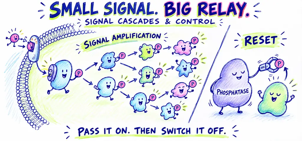
A signal transduction cascade relays information through a cell. Some steps amplify the input, branches connect it to different responses, and termination mechanisms limit its duration. To analyse a disrupted pathway, locate the affected step and predict what changes upstream and downstream.
A frightened animal can mobilize fuel rapidly after an endocrine signal. A small number of activated receptors can change the activity of many downstream molecules. That multiplication is signal amplification.
Watch the process
Signal Transduction Pathways
Not every step amplifies, and every real pathway has limits. The models below use specified branching factors to make the arithmetic clear. Signal termination, available substrates, and feedback determine the response in an actual cell.
Following One Pathway All the Way Through
Follow epinephrine signaling in a liver cell from the receptor to glucose release. At each step, identify the physical change: nucleotide binding, enzyme activation, messenger production, or phosphorylation.
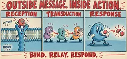
1. Reception: Epinephrine, which is hydrophilic, binds the extracellular face of a G protein-coupled receptor. The message is now a receptor shape.
2. G protein activation: The shape change lets the receptor act on the associated G protein, which releases GDP and binds GTP. The message is now the identity of a nucleotide bound to a protein.
3. Enzyme activation: The GTP-bound G protein subunit detaches, diffuses in the plane of the membrane, and binds adenylyl cyclase, the membrane enzyme that makes cyclic AMP from ATP. The message is now an enzyme’s catalytic activity.
4. Second messenger: Adenylyl cyclase converts many ATP molecules into cyclic AMP, which diffuses through the cytosol. The message is now a concentration.
5. Kinase activation: Cyclic AMP binds the regulatory subunits of protein kinase A, releasing its catalytic subunits. The message is a second enzyme activity.
6. Cascade: Protein kinase A phosphorylates and activates phosphorylase kinase, and phosphorylase kinase in turn phosphorylates and activates glycogen phosphorylase. The message is a chain of covalent modifications.
7. Response: Glycogen phosphorylase releases mainly glucose-1-phosphate from glycogen. Liver cells convert this through glucose-6-phosphate to free glucose that can enter the blood. The message has become a metabolic output.
Seven stages, and the hormone participated only in the first one. Each arrow is a conversion between physical forms, which is what the word transduction means.
The Second Major Membrane Route: Calcium and IP\(_3\)
Not every GPCR talks to adenylyl cyclase. A large family activates phospholipase C instead, an enzyme that cleaves a particular membrane phospholipid into two fragments that both act as messengers. One fragment, IP\(_3\) (inositol trisphosphate), is water soluble and diffuses to the endoplasmic reticulum, where it opens calcium channels. The other fragment, DAG (diacylglycerol), stays in the membrane and helps activate protein kinase C.
The calcium arm is the one to understand mechanically. Free calcium concentration is normally much lower in the cytosol than in the ER or outside the cell. Channels let calcium move down its electrochemical gradient; transport systems maintain or restore that gradient. Calcium signals can change quickly, but their size and timing vary by cell and stimulus. Calcium participates in processes such as muscle contraction and neurotransmitter release.
Kinase Cascades and What They Buy
One common pathway downstream of a receptor tyrosine kinase uses a MAP kinase cascade: a MAP kinase kinase kinase activates a MAP kinase kinase, which activates a MAP kinase. The final kinase can act on cytoplasmic targets or regulate nuclear transcription factors. This provides one route from a surface growth-factor signal to altered gene expression.
A kinase cascade can amplify a signal because each active kinase modifies many copies of the next. Its layers also provide separate sites for regulation. With suitable enzyme properties and feedback, the pathway can show a threshold response; amplification alone does not guarantee that behavior.
Calculate Amplification in a Hypothetical Model
In this hypothetical model, a single growth-factor molecule binds one receptor. That receptor activates 8 molecules of the first kinase in a cascade. Each active first kinase phosphorylates 30 molecules of the second kinase. Each active second kinase phosphorylates 40 molecules of the third kinase. Each active third kinase phosphorylates 25 transcription factor molecules. Assume distinct targets, sufficient ATP and substrate, and negligible deactivation during the stated interval. Find the number of activated transcription factors, and then find the amplification factor of the cascade stages alone, excluding the receptor step.
Multiply outward, one layer at a time, and label the units as you go so nothing gets lost. \[1 \times 8 = 8 \text{ first kinases}, \qquad 8 \times 30 = 240 \text{ second kinases}\] \[240 \times 40 = 9600 \text{ third kinases}, \qquad 9600 \times 25 = 240{,}000 \text{ transcription factors}\] So one binding event activates \(2.4 \times 10^{5}\) transcription factors.
For the second part, read the question carefully: it asks for the amplification of the cascade stages only, which means starting the count at the 8 first kinases rather than at the single receptor. Divide the output by that starting number: \[\frac{240{,}000}{8} = 30{,}000 .\] You could also get this by multiplying only the per-stage branching factors that lie inside the cascade, \(30 \times 40 \times 25 = 30{,}000\), which is a good check: the amplification factor of a series of stages is the product of the individual branching factors, independent of how many molecules entered.
Now interpret the number with its units: the supplied model yields 30,000 activated transcription factors per initially active first kinase. This count cannot be converted directly into an equivalent extracellular hormone concentration. Binding affinities, compartment volumes, time, and substrate limits would also matter. A constitutively active kinase can drive downstream signaling without the usual input, but the magnitude depends on those conditions.
Answer
\(2.4 \times 10^{5}\) activated transcription factors; the cascade stages amplify \(30{,}000\)-fold, the product of the per-stage factors.
Turning the Pathway Off Is an Active Job
A response ends when activating signals are removed or the active components are inactivated. Follow four termination mechanisms in this pathway.
The ligand dissociates. For a reversibly bound ligand, falling ligand concentration can reduce receptor occupancy. Receptor regulation and the lifetime of active downstream components also influence how quickly the response ends.
The G protein hydrolyzes its own GTP to GDP. This is a built-in timer: the same protein that carries the message destroys the message at a rate that depends on the protein and its regulators. The bacterial toxin that causes cholera works by blocking exactly this step in intestinal cells, so the G protein stays on, cyclic AMP stays high, and the cells maintain elevated chloride secretion through channels. Water follows the resulting osmotic gradient; cells do not pump water directly.
Phosphodiesterase converts cyclic AMP into AMP, so the second messenger disappears once production stops. Calcium signaling ends by a different mechanism: transport systems move calcium back into the ER or out of the cell.
Protein phosphatases remove the phosphates that kinases added. This is the step that makes a phosphorylation cascade a switch rather than a one-way trip. A cell contains both kinases and phosphatases acting on the same targets continuously, and the phosphorylation state of any protein is the balance between the two rates, not the result of a single event.
Where Did the Signal Break?
A cell line responds to a hormone by raising cyclic AMP and activating protein kinase A. Three treatments are tested. Use a linear model in which this receptor is the only hormone input, protein kinase A is required for the measured response, and compensating feedback is negligible. In each case, decide what happens to cyclic AMP concentration and the response.
Treatment 1: a drug that blocks the hormone’s binding site on the receptor. Reception fails, so no G protein is activated, adenylyl cyclase is never switched on, cyclic AMP stays at baseline, and the response does not occur. Nothing downstream is broken, which matters: delivering cyclic AMP into the cytosol, or using a suitable membrane-permeant analog, should rescue the response. Rescue supports functional downstream machinery; by itself it localizes a defect upstream of kinase activation, not uniquely to reception.
Treatment 2: a drug that inhibits phosphodiesterase. Removal of cyclic AMP is reduced while its production remains intact. With ongoing production, cyclic AMP can accumulate; after a hormone pulse, it can remain elevated longer. The response can therefore be prolonged. Its exact magnitude and duration depend on residual removal and other regulatory mechanisms. Ask yourself which direction the defect points: in this model, inhibiting removal prolongs the messenger signal.
Treatment 3: a mutation in the regulatory subunits of protein kinase A that prevents cyclic AMP binding. Reception, the G protein, adenylyl cyclase, and cyclic AMP production are all untouched, so cyclic AMP rises normally, but the measured phosphorylation response still fails. The kinase cannot be released by its regulatory subunits, so the hormone-dependent phosphorylation of these target proteins fails and the measured response is absent. Other kinases can still phosphorylate other proteins. A normal second messenger concentration is evidence about the upstream half of the pathway only.
Answer
Blocking reception gives low cyclic AMP and no response; blocking phosphodiesterase gives high, prolonged cyclic AMP and a prolonged response; a PKA regulatory-subunit defect that prevents cyclic AMP binding gives normal cyclic AMP but no measured PKA-dependent response in this model.
One Signal, Several Branches
A receptor tyrosine kinase, once cross-phosphorylated, presents a cluster of docking sites, and different relay proteins bind different sites. One ligand binding event can therefore launch a cell-division pathway, a survival pathway, and a metabolic pathway simultaneously from the same receptor. Branching changes how a pathway responds to a selective disruption.
First, a selective block in one independent branch can leave another functional. If division is inhibited but a survival response remains, a branch-specific defect is consistent with the evidence. Partial receptor inhibition, different response thresholds, or connections between branches are alternatives unless the experiment excludes them.
Second, the same second messenger can produce opposite outcomes in two cell types, because what a cell does with elevated calcium or cyclic AMP depends on which substrate proteins that cell owns. Cyclic AMP triggers glycogen breakdown in a liver cell and hormone secretion in an endocrine cell. Receptor context, effector proteins, substrates and regulatory interactions help determine the response to the messenger.
Review: Inside the Pathway: Cascades, Branching, and Shutting Off
A transduction pathway converts information through connected components; some steps amplify, and regulatory mechanisms terminate or limit the response.
Disabling a required activation step can reduce the response; disabling termination can prolong it. Use the pathway and conditions shown.
Do not treat a measured second messenger level as proof the whole pathway works. It reports only on the steps above the measurement.
Distinctions to keep clear
Distinguish amplification per input from total response. In a simplified branching calculation, each initiating molecule produces the number of outputs specified by the multiplying factors. More initiating molecules then produce more total output. In living cells, saturation, limited substrates, signal duration, and feedback can change those factors. A constant amplification factor is a model assumption, not a universal biological law.
Kinases and phosphatases change phosphorylation in opposite directions. Both are enzymes that act on the same target proteins, and their names sound similar. A kinase adds a phosphate group taken from ATP, generally activating or inactivating the target by changing its shape. A phosphatase removes that phosphate; the effect on activity depends on the target and phosphorylation site. If an exam stem inhibits one of them, decide first which direction the target’s phosphorylation state will drift: if competing reactions continue, inhibiting the kinase tends to reduce target phosphorylation, while inhibiting the phosphatase tends to increase it. This does not by itself tell you whether the target becomes more active or less active.
Transduction pathways and their off-switches
Practice question 1
Cholera toxin modifies a G protein so that it can no longer hydrolyze bound GTP. In an intestinal cell that uses this G protein, the most direct consequence is that
-
the G protein returns more rapidly to its inactive GDP-bound state
-
adenylyl cyclase remains activated and cyclic AMP stays elevated even without ligand
-
cyclic AMP declines because the G protein cannot complete its normal off-switch
-
adenylyl cyclase becomes inactive as soon as ligand dissociates, despite the G protein remaining active
Practice question 2
A cell is treated with a hormone and its cyclic AMP concentration rises normally, but the usual phosphorylation of target proteins does not occur. The defect is most likely in
-
the receptor
-
adenylyl cyclase
-
protein kinase A
-
phosphodiesterase
Practice question 3
In a model, one active kinase activates 20 second kinases; each second kinase activates 20 third kinases; each third kinase activates 20 distinct final targets. Assume sufficient substrate and no deactivation. Relative to one initiating kinase, this cascade
-
activates 60 final targets because the three branching factors are added
-
activates 8000 final targets and provides several steps at which activity can be regulated
-
activates 400 final targets because only kinase-to-kinase steps count toward the final output
-
activates 20 final targets because only the last kinase layer contributes to amplification
Practice answer key
1. B; 2. C; 3. B.
Practice answer explanations
-
Transduction pathways and their off-switches, Question 1. Choice B is correct. Preventing GTP hydrolysis prolongs the active G-protein state and stimulation of adenylyl cyclase in this pathway. Choice A reverses the nucleotide-state effect. Choice C mistakes failure of termination for failure of activation. Choice D assumes ligand removal can shut off the downstream G protein even when its own off-switch is disabled.
-
Transduction pathways and their off-switches, Question 2. Choice C is correct. The normal hormone-induced cyclic AMP rise supports functional upstream signal production. Among the choices, impaired protein kinase A best explains failure of the usual target phosphorylation. Choices A and B would more naturally reduce hormone-induced cyclic AMP production. Loss of phosphodiesterase in D would prolong cyclic AMP rather than prevent kinase activation. These data support a location; they do not prove every upstream component is completely normal.
-
Transduction pathways and their off-switches, Question 3. Choice B is correct. The specified model gives \(20\times20\times20=8000\) final targets per initiating kinase. Choice A adds sequential multiplying factors. Choice C stops at the third-kinase count and omits its target activation. Choice D ignores amplification before the final step. Distinct regulatory proteins can act at the different cascade stages.
Continue your review at the AP Biology study hub.

Related to This Article
More math articles
- Hypotheses and Judging a Good Experiment
- Communication, Service Recovery, HIPAA, and DEI
- FTCE Mathematics 6-12 Flashcards
- Signing statements
- FREE 4th Grade Common Core Math Practice Test
- Economic cost includes implicit opportunity cost
- How to Avoid Careless Errors in Standardized Math Tests
- Business groups
- FREE 8th Grade OST Math Practice Test
- PERT Math – Test Day Tips






What people say about "Signal Transduction Cascades: Amplification and Control"?
No one replied yet.