Cyclins and CDKs: How They Control the Cell Cycle
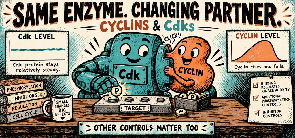
Cyclins are regulatory proteins that bind cyclin-dependent kinases (CDKs). CDKs are enzymes that phosphorylate target proteins. Changing cyclin levels helps control when these complexes form, while phosphorylation and inhibitors adjust their activity.
Early frog embryos provide a way to study repeated cell-cycle transitions. Researchers can prepare egg cytoplasm extracts that reproduce cycles of mitotic activity outside an intact embryo. Changing cyclin synthesis or degradation changes those cycles.
Watch the process
Cell cycle control | Cells | MCAT | Khan Academy
These experiments support the inference that a molecular control system in the cytoplasm can generate oscillating activity. Early embryonic cycles use stored material and differ from ordinary growing somatic cells, so their timing is not a universal cell-cycle schedule.
Why Two Proteins Instead of One
A cyclin-dependent kinase, or Cdk, is an enzyme that transfers phosphate groups to other proteins and, by doing so, changes what those proteins do. In the model used here, total Cdk protein stays relatively stable through the cycle. For the cell-cycle Cdks in this model, cyclin binding is necessary for normal kinase activity.
A cyclin is a regulatory protein that has no catalytic activity of its own and whose concentration rises and falls in a repeating pattern each cycle, which is where the name comes from. Cyclin binding helps arrange the Cdk active site for catalysis; phosphorylation and inhibitor proteins provide additional controls. It also helps steer the kinase to the right substrates, so different cyclins bound to the same Cdk send it after different targets.
This arrangement permits activity to change without repeatedly making and destroying the Cdk protein itself. Cyclin synthesis and degradation change the availability of active complexes, while other regulators adjust their activity. Both protein production and protein modification therefore help control timing.
What the Cyclin Curve Shows
Consider a hypothetical experiment with a synchronized population of sea urchin embryos, in which most cells are at a similar point in the cycle. Samples are taken every 10 minutes across two division cycles, and two things are measured in each sample: the amount of cyclin protein, and the kinase activity of the cyclin-Cdk complex. Cyclin rises steadily from a low point, peaks, then drops almost to zero within a few minutes. Kinase activity stays near zero while cyclin is low and rising, then rises sharply once cyclin is well past the midpoint of its climb, peaks just after cyclin does, and collapses with it. Cdk protein is flat across the whole record. What can be concluded, and what cannot?
Claim. The measurements are consistent with cyclin-dependent regulation and rapid cyclin removal at mitotic exit.
Evidence. Cdk protein is flat while activity oscillates, so activity is not set by how much kinase there is. Activity tracks cyclin, but not proportionally: it stays near zero through the early part of cyclin’s rise and then climbs steeply. Cyclin falls to near zero in a few minutes, far faster than it rose.
Reasoning. A flat protein level with oscillating activity shows that changing total kinase abundance is not the explanation for its changing activity. A binding partner or chemical modification could regulate it. The delayed rise in kinase activity is consistent with switch-like regulation, but this time course alone does not identify a unique cyclin threshold or establish the mechanism. Controlled cyclin-dose experiments can test that hypothesis. The speed of the fall is the strongest single clue in the data. Dilution by growth cannot remove a protein in minutes, and neither can simply shutting off transcription, because existing protein would remain. Rapid degradation is a strong candidate, although these concentration measurements alone do not establish the removal mechanism.
What cannot be concluded. These data show correlation between cyclin level and kinase activity across time. They do not by themselves prove that cyclin binding causes activation, since some third factor could rise and fall on the same schedule. Adding purified cyclin to a controlled extract can test activation, and preventing cyclin degradation can test its role in mitotic exit. Appropriate untreated controls and measurements of the relevant proteins distinguish these explanations.
Conclusion
Activity changes without changing total Cdk, and its pattern is consistent with threshold regulation by cyclin. The abrupt cyclin decline supports testing regulated degradation.
The G\(_2\) Oscillator in Detail
The mitotic cyclin-Cdk complex is often called MPF, for maturation-promoting factor, a name left over from the frog experiments. Follow one turn.
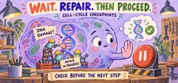
In a simplified model of a growing cell, mitotic cyclin accumulates during S phase and G\(_2\). Inhibitory phosphorylation can keep cyclin-Cdk complexes inactive. A phosphatase called Cdc25 removes inhibitory phosphates, while feedback can make the rise in activity abrupt. DNA replication and damage checkpoints can delay this activation. Cyclin concentration alone does not tell you whether all these controls have been satisfied.
Active MPF then phosphorylates a specific set of targets, and every one of them maps onto something you can see under a microscope. Phosphorylating the nuclear lamins disassembles the nuclear envelope. Phosphorylating condensin proteins condenses the chromosomes. Phosphorylating microtubule-associated proteins reorganizes the cytoskeleton into a spindle. The abstract statement “MPF triggers mitosis” becomes a list of concrete events with named substrates.
Protein Destruction at Anaphase
The metaphase-to-anaphase transition is regulated differently, and it is the sharpest example of protein destruction as a control mechanism.
Sister chromatids are held together by a protein ring, and that ring is cut by a protease, an enzyme that cuts other proteins. The protease is kept inactive by an inhibitor bound to it. A large complex called the anaphase-promoting complex, or APC, tags that inhibitor for destruction. The moment the inhibitor is destroyed, the protease cuts, the rings fall apart, allowing sister chromatids to separate during anaphase.
The spindle assembly checkpoint restrains the Cdc20-activated form of the APC. An unattached kinetochore generates a diffusible inhibitory signal, and one unattached kinetochore anywhere in the cell is enough to delay APC-dependent anaphase. This explains why one badly attached chromosome can stop a whole cell. The inhibitory signal restrains APC activity throughout the cell.
The APC does one more thing. It also tags mitotic cyclin for destruction, which is what drops Cdk activity at the end of mitosis and allows phosphatases to reverse many mitotic phosphorylation events. Chromosomes decondense, envelopes reform, and the cell returns to G\(_1\). Anaphase and mitotic exit are therefore driven by the same machine acting on two different targets, so blocking cyclin destruction can prevent mitotic exit even when sister chromatids have separated.
The Restriction Point and the Brakes on It
The G\(_1\) decision is the one connected to the outside world, and it is where growth signaling connects to cell-cycle control.
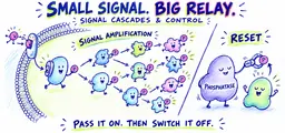
A growth factor binds a receptor tyrosine kinase, the transduction pathway runs a kinase cascade to the nucleus, and transcription factors switch on the gene for a G\(_1\) cyclin. G\(_1\) cyclin binds its Cdk. The complex phosphorylates a tumor-suppressor protein called Rb, which in an unphosphorylated state is clamped onto transcription factors and keeps them silent. Phosphorylating Rb releases those transcription factors, they switch on the genes for DNA replication enzymes and for S-phase cyclins, and the cell crosses the restriction point.
Rb restrains the cycle by binding transcription factors needed for S phase. Growth signaling can release that restraint through Rb phosphorylation. Losing Rb can bypass this control, although other controls and growth requirements remain.
Two kinds of brake act on the cyclin-Cdk complexes themselves. A Cdk inhibitor protein binds an assembled cyclin-Cdk complex and shuts off its kinase activity directly. The best-known one is produced when the p53 protein, which accumulates in response to DNA damage, activates transcription of its gene. And when damage is too severe to repair, p53 instead drives the expression of genes that commit the cell to apoptosis, a controlled process of cellular dismantling, in which the cell fragments itself into membrane-bounded packages that neighbors clear away usually without the inflammatory response associated with uncontrolled cell rupture.
Three Ways to Make a Cell Divide Without Permission
A researcher generates three cell lines, each with a single change, and finds that all three divide in medium containing no growth factor, which normal cells cannot do. Line 1 expresses a G\(_1\) cyclin continuously from an artificial promoter. Line 2 has lost both copies of the Rb gene. Line 3 carries a receptor tyrosine kinase locked in the dimerized state. Use the simplified receptor-to-cyclin-to-Rb pathway described here, with other growth requirements supplied. Explain each and rank how far down the pathway the lesion sits.
Work from the shared requirement. Normally, growth factor is needed because, in this model, it normally promotes enough G\(_1\) cyclin production to phosphorylate Rb and release the S-phase transcription factors. Anything that supplies that outcome by another route removes the requirement.
Line 3 is the most upstream lesion. A permanently dimerized receptor cross-phosphorylates with no ligand present, so the cascade runs, the transcription factors that induce cyclin are activated, and cyclin appears. Everything downstream of the receptor is normal and is simply being driven by a false input. This cell would still respond to a drug that inhibits the kinase cascade.
Line 1 sits below that. The cascade is irrelevant here, because the cyclin is supplied directly. A drug that blocks the receptor or the cascade would not restore control, which is a useful experimental signature: the level at which a treatment stops working tells you where the lesion is.
Line 2 is the most downstream of the three, and also the most complete. Without Rb, the S-phase transcription factors are free regardless of cyclin, so this Rb-dependent transcriptional brake is released. Other cyclin-Cdk functions remain necessary for DNA replication and division. Restoring functional Rb is one way to test this model; blocking a required downstream process can still prevent division.
The ranking is the answer to the deeper question of why cancers carry mutations at many different steps: changes at different positions can bypass the same control, and an upstream inhibitor may fail to reverse a downstream defect. This does not rank all possible cancer treatments.
Answer
All three bypass the growth-factor requirement, at the receptor (Line 3), at cyclin supply (Line 1), and at the Rb brake itself (Line 2); the more downstream the lesion, the less any upstream intervention can correct it.
Review: Cyclins, Cdks, and the Machinery Behind the Checkpoints
Cell-cycle transitions are made by regulatory complexes that are switched on by protein accumulation and switched off by protein destruction.
Change what is made or what is destroyed and you change when, or whether, a transition happens.
In this simplified model, total Cdk abundance stays relatively stable while cyclin abundance changes. Cyclin degradation reduces the amount of active complex, and phosphorylation and inhibitor proteins provide additional control. More Cdk protein alone does not establish more cell division.
Distinctions to keep clear
A checkpoint is a regulatory system, not the protein complex it controls. Students write that the G\(_2\) checkpoint “is” MPF, or that the spindle assembly checkpoint “is” the APC. They are not the same thing. MPF and the APC are the engines that drive transitions forward. The checkpoints are regulatory systems that hold those engines off until conditions are met. Loss of a checkpoint can permit inappropriate progression, although other controls, missing resources, or severe damage may still prevent division.
Whether increased activity or lost function promotes cancer depends on the protein’s normal role. Cyclins and their Cdks push the cycle forward, so a mutation that raises their activity is dangerous, and one activating allele can be sufficient to alter that pathway; it need not be sufficient to cause cancer. Rb, p53, and the Cdk inhibitors hold the cycle back, so losing their function can remove a restraint on proliferation. Two-hit loss is an important tumor-suppressor model, but it is not a universal rule for every regulator or mutation.
Cyclin-Cdk control and checkpoint machinery
Practice question 1
In a synchronized cell population, Cdk protein concentration is constant across the cycle while mitotic cyclin concentration rises through S and G\(_2\) and falls abruptly during mitosis. Cdk activity tracks cyclin, and blocking protein degradation prevents the cyclin decline. These data best support the conclusion that
-
Cdk activity is limited by cyclin availability, and cyclin is removed by regulated destruction rather than by dilution
-
Cdk is synthesized once per cycle and degraded at the end of mitosis
-
cyclin is a kinase that phosphorylates Cdk to inactivate it
-
cyclin and Cdk act in separate pathways that never interact
Practice question 2
A mutation prevents the anaphase-promoting complex from tagging the inhibitor of the protease that separates sister chromatids. Cells carrying this mutation would most likely
-
fail to replicate their DNA and arrest in G\(_1\)
-
condense chromosomes and align them at the metaphase plate but never begin anaphase
-
enter mitosis without a spindle
-
exit mitosis before chromosomes condense
Practice question 3
Cells that have lost both functional copies of the Rb gene divide in medium containing no growth factor. The best explanation is that Rb normally
-
transports growth factors into the nucleus
-
binds and sequesters transcription factors needed for S phase until a growth signal causes it to be phosphorylated
-
degrades G\(_1\) cyclin so that Cdk stays inactive
-
attaches kinetochores to spindle microtubules during metaphase
Practice answer key
1. A; 2. B; 3. B.
Practice answer explanations
-
Cyclin-Cdk control and checkpoint machinery, Question 1. Choice A is correct. Constant Cdk abundance with activity tracking cyclin supports cyclin-dependent regulation. The added degradation-block experiment directly supports protein destruction as the cause of the cyclin decline. Choice B contradicts constant Cdk abundance. Choice C reverses the catalytic roles. Choice D contradicts the coupled behavior and the known cyclin-Cdk complex.
-
Cyclin-Cdk control and checkpoint machinery, Question 2. Choice B is correct. The anaphase-promoting complex triggers anaphase by marking for destruction the inhibitor of the protease that cuts the link between sister chromatids, so blocking that step leaves chromosomes condensed and aligned but unseparated in this simplified model. Choice A names a phase governed by an entirely different complex. Choice C describes spindle assembly, which depends on mitotic Cdk activity rather than on the anaphase-promoting complex. Choice D reverses the order of events within mitosis.
-
Cyclin-Cdk control and checkpoint machinery, Question 3. Choice B is correct. Rb holds S-phase transcription factors inactive until a growth signal produces enough G\(_1\) cyclin-Cdk activity to phosphorylate it, so losing Rb frees those factors and explains the growth-factor-independent behavior specified in this model; other growth requirements still matter. Choice A assigns Rb a transport role it does not have. Choice C reverses the relationship, since Rb is a target of cyclin-Cdk rather than a destroyer of cyclin. Choice D confuses Rb with the spindle assembly machinery.
Continue your review at the AP Biology study hub.

Related to This Article
More math articles
- Distinguishing correlation from causation
- Algebra 1 Fundamentals: A Guide for Students and Teachers
- Point-Slope Form Worksheet with Answers
- Full-Length 7th Grade GMAS Math Practice Test-Answers and Explanations
- The Best Grade 5 ELA Practice Tests for Minnesota Students
- Math in Everyday Life: How to See the World Through Numbers
- 10 Most Common 5th Grade PSSA Math Questions
- How to Convert, Compare, Add, and Subtract Mixed Customary Units?
- Kennedy’s New Frontier and Cold War Development Policy
- GED Calculator Guide: Learn How To Use The TI-30XS







What people say about "Cyclins and CDKs: Cell-Cycle Control Explained"?
No one replied yet.